I was recently referred a patient who started to complain of a purple spot in the vision of the right eye. The patient is 74 years old and had just received an injection of Botox and some dermal fillers for treatment of facial wrinkles.
After the patient arrived at my office, a complete dilated eye exam was performed and I determined that my patient had developed a branch retinal artery occlusion (BRAO). There are two types of artery occlusions; central retinal artery occlusion (CRAO) which involves the entire retina and branch retinal artery occlusion (BRAO) which involves a portion of the retina.
Branch Retinal Artery Occlusions
There are two general classes of retinal vascular occlusions. There are vascular occlusions (blockage of blood flow) of the retinal veins and vascular occlusions of the retinal arteries.
Artery occlusions occur after a blood vessel is completely blocked by an object, called an embolus. An embolus is usually cholesterol, but can also be blood clots or fat. Retinal vein occlusions are not blocked by an embolus, but rather are squeezed close by an adjacent rigid blood vessel.
In addition to my examination, a fluorescein angiogram was performed to help make the diagnosis. Both artery and venous occlusions have characteristic findings on the fluorescein angiogram.
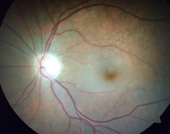
Hollenhorst Plaque and Retinal Edema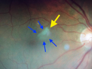
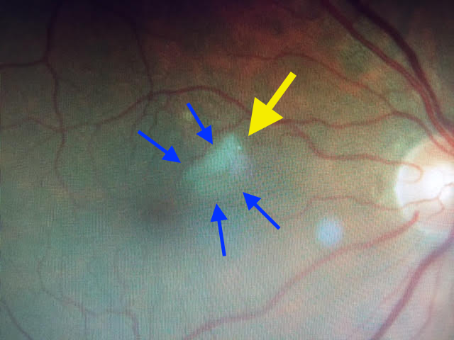
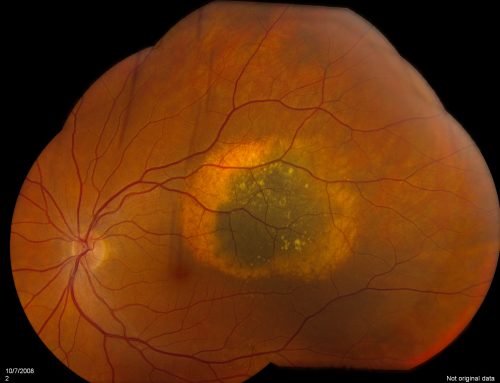
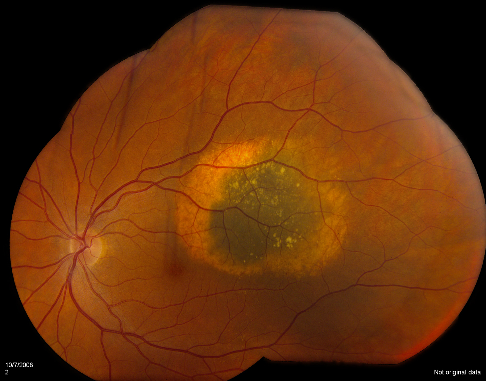
This is a picture of the retina of the right eye of my patient. The yellow arrow shows a bright white speck of cholesterol which is blocking a very tiny retinal artery. The blue arrows highlight the retinal edema, or swelling, caused by the blockage.
Click on the image to get a larger view.
Hollenhorst plaques are characteristic of artery occlusions and can be clearly seen to cause a blockage.
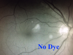
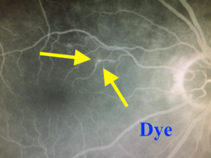
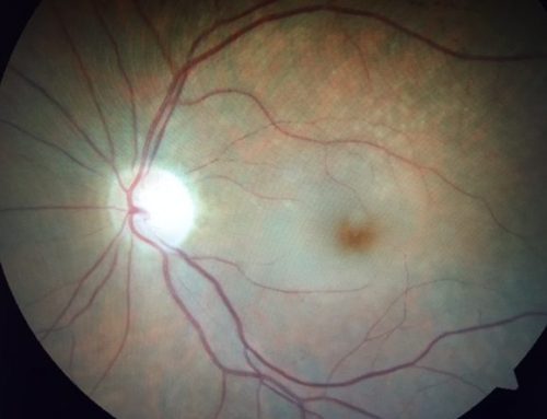
Fluorescein Angiogram
A fluorescein angiogram is a diagnostic test requiring injection of a vegetable based dye into the bloodstream. The dye travels to the retina and becomes visible when using a special filter. This is a great way to visualize blood flow. The two pictures nicely demonstrate “with” dye and “without.” The dye appears white and represents the blood flow.
Risk of Stroke
In most cases, the vision loss from artery occlusions is permanent. There can be complications that arise from the artery occlusions so follow up with a retina specialist remains important.
The patient might have had a stroke had the plaque traveled to the brain. In cases of artery occlusions, it is important to assess the patient’s risk of developing a stroke. This requires team approach with the patient’s family doctor and possibly cardiologist.

