As a retina specialist in Orange County, CA, patients are often referred to me with sudden loss of vision. This is an example of the importance of an eye examination.
A 25 year old healthy man was referred to me last week because of sudden vision loss to his right eye.
His vision was worse than 20/400, that is, he could not see the big “E” on the eye chart. Sudden vision loss like this usually is due to a problem in the retina, optic nerve or even the brain. Retinal detachment, bleeding, loss of blood flow can cause sudden vision loss, but in a healthy young man?
I needed to dilate his pupils and examine his retinas. After I dilated the pupils, I saw multiple areas of bleeding in both eyes (see images below), which is unusual for a person this young without any medical problems.
Retinal Photos and Fluorescein Angiogram
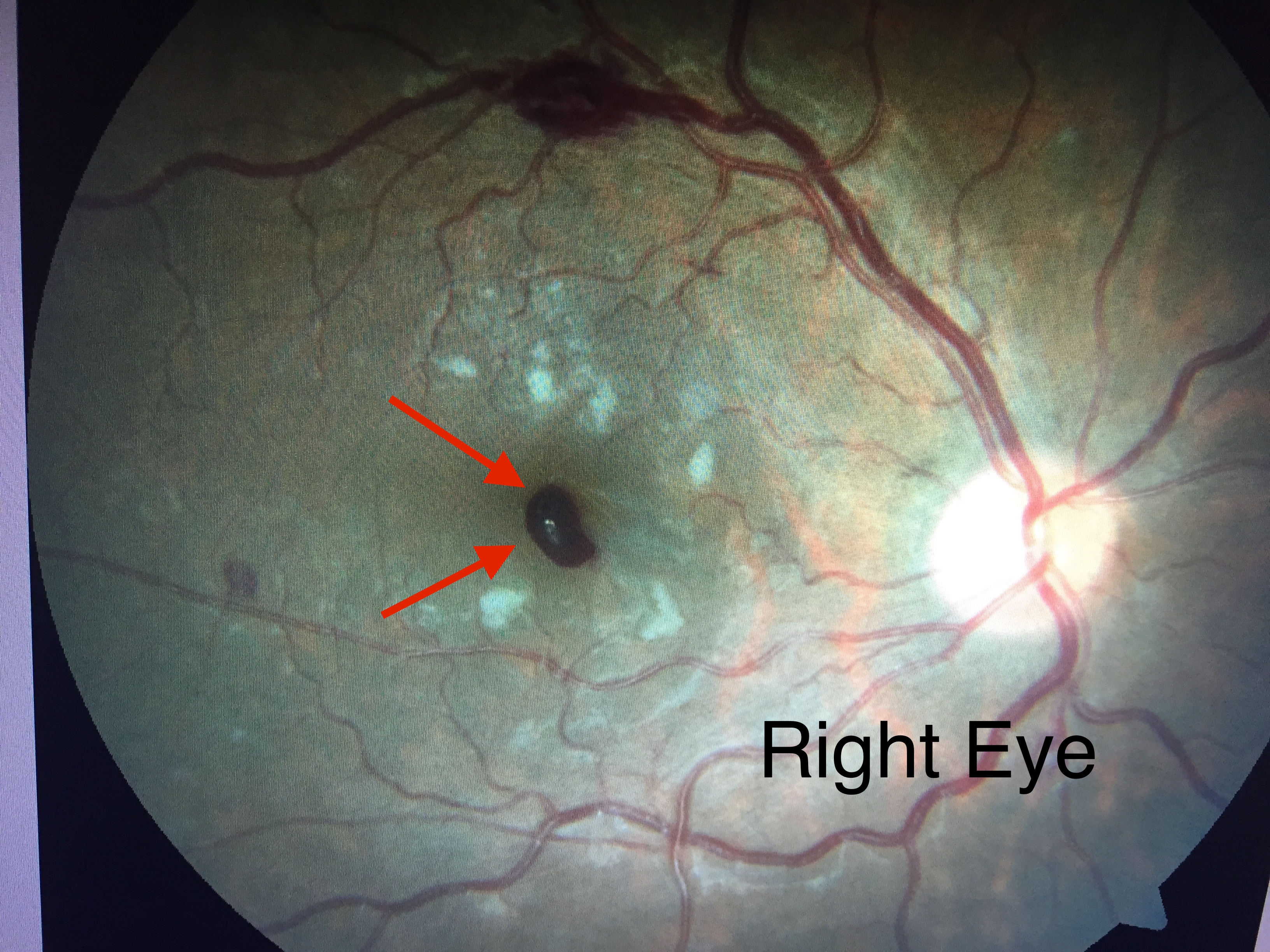
 This is a picture of the patients right eye. Note the blood highlighted by the red arrows. This “preretinal hemorrhage” is lying in front of the macula, thus blocking his vision.
The white spots are known as soft exudates and are due to a circulation/oxygenation problem causing the nerves of the retina to turn white.
The blood at the top, while abnormal, is not affecting his vision.
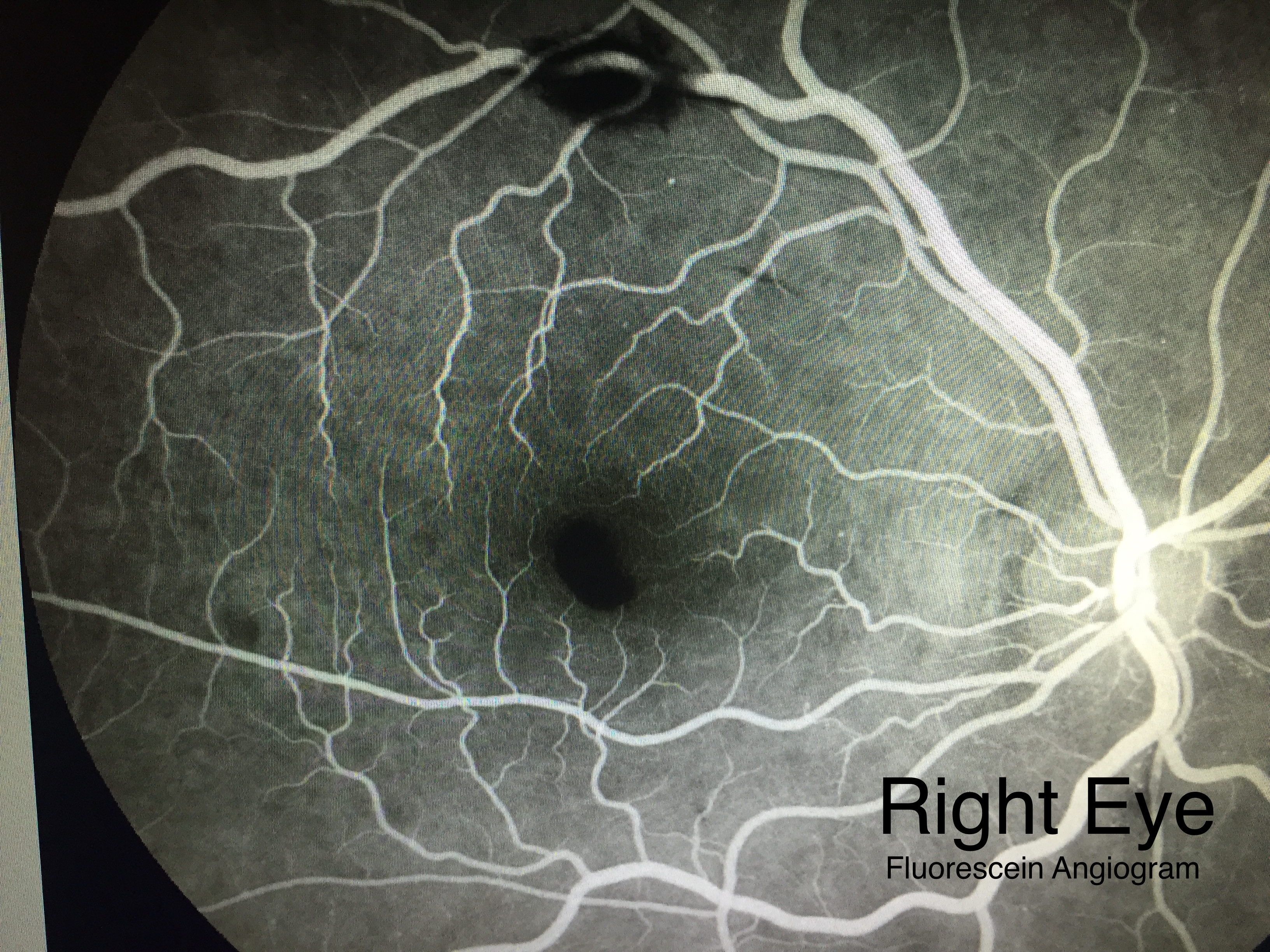 A fluorescein angiogram is often used by a retina specialist to examine the health and integrity of the retina, especially blood flow.
If you look carefully (click the picture to enlarge), the kidney shaped hemorrhage can still be seen in this fluorescein angiogram, proving that block effect on the vision.
The macula is normally darker making it more difficult to see the blocking effect of the blood.
 This is the picture of the patients left eye. Note that the macula (arrows) is free of blood, hence explaining the 20/20 vision.
Similar soft exudates are seen. There is also blood near the optic nerve and to the side of the macula.
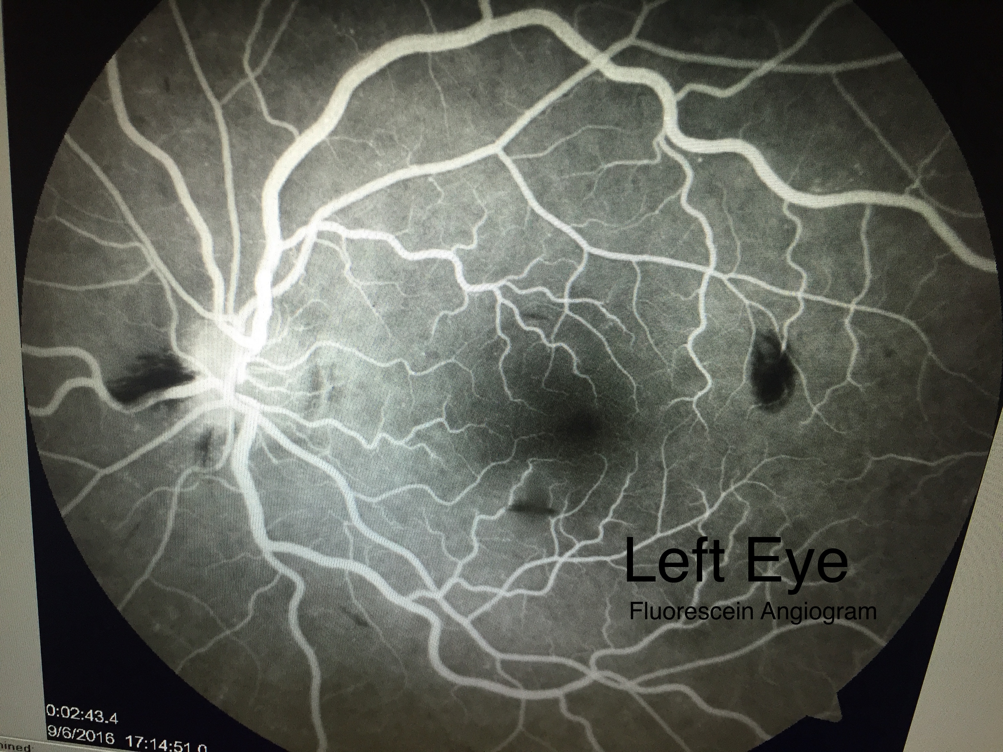 The fluorescein angiogram of the left eye.
It is important to note that the macula is free of blood and is normal in this study.
The dark areas are blood, away from the macula and do not affect the vision.
Symptoms of Fatigue
After further questioning, he admitted to feeling fatigued over the past 2 months, and occasionally getting short of breath when climbing stairs. Again, this is very unusual especially since my patient states he exercises regularly.
Based on the above combination of symptoms and retinal exam findings, I suspected that he had a blood disorder, so I sent him for urgent blood work. Unfortunately, this test showed that the patient was in the acute phase of leukemia. He is now admitted at a local hospital and undergoing chemotherapy.
Prognosis
Fortunately, my patient’s vision is likely to improve and return without treatment. Blood of this nature is quite benign and should absorb by itself. As the premacular hemorrhage gets smaller, the vision should improve.
The bleeding has occurred because his platelet count was abnormally low from the leukemia. His white blood count was 4-5 times normal making his blood thicker and causing oxygenation problems.
The fluorescein angiogram was essentially normal which allows me to be so confident about his visual prognosis.
It’s too early to make a statement about the leukemia, but it is fortunate that the bleeding caused the acute loss of vision. Had he not bled, it is likely he would not be receiving treatment at this time.
If you have any questions or concerns, please leave a comment below. If you live in or near the Orange County, CA area, please call us.